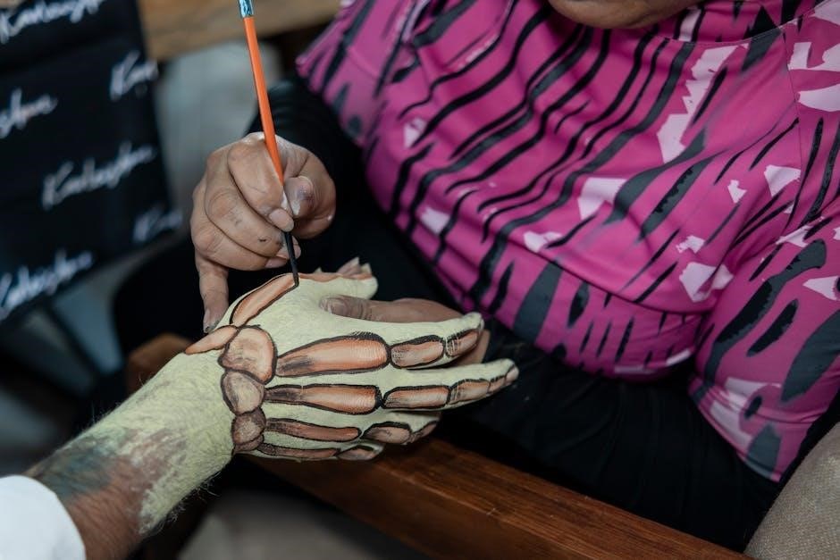
The adult human body contains 206 bones, forming a complex framework essential for movement, support, and protection. This skeletal system is divided into axial and appendicular bones, each serving unique functions. Understanding the structure and arrangement of these bones is crucial for appreciating human anatomy and health. A detailed PDF diagram of the 206 bones provides a visual guide, helping learners identify and study each bone’s location and role in the body.
1.1 Overview of the Skeletal System
The human skeletal system consists of 206 bones, forming a framework that provides support, protection, and facilitates movement. It is divided into the axial skeleton (80 bones) and the appendicular skeleton (126 bones); The axial skeleton includes the skull, vertebral column, and ribcage, while the appendicular skeleton comprises the limbs and pelvis. This system is essential for overall bodily functions and stability. A detailed PDF diagram of the 206 bones offers a visual understanding of their structure and arrangement.
1.2 Importance of Understanding the 206 Bones
Understanding the 206 bones is vital for comprehending human anatomy and health. Each bone plays a unique role, from supporting the body to facilitating movement. Knowledge of bone structure aids in diagnosing injuries, preventing conditions like osteoporosis, and appreciating the skeletal system’s role in overall well-being. A PDF diagram of the 206 bones provides a clear, educational resource for study and reference.
Classification of Bones
The 206 bones are classified into two main categories: the axial skeleton and the appendicular skeleton. The axial skeleton includes bones of the skull, spine, and ribcage, while the appendicular skeleton comprises the bones of the limbs and pelvis. This classification helps in understanding the structural and functional organization of the skeletal system, as detailed in a PDF diagram.
2.1 Axial Skeleton (80 Bones)
The axial skeleton consists of 80 bones forming the body’s central framework. It includes the skull, vertebral column, ribcage, and sternum. These bones protect vital organs like the brain, heart, and lungs, while providing structural support and attachment points for muscles. A PDF diagram of the axial skeleton helps identify its components, such as the cranium, face bones, vertebrae, and ribs, highlighting their roles in maintaining posture and facilitating movement.
2.2 Appendicular Skeleton (126 Bones)
The appendicular skeleton comprises 126 bones, primarily forming the upper and lower limbs, pelvis, and shoulder girdle. It enables movement, stability, and weight distribution. This skeleton includes the scapula, humerus, radius, ulna, carpals, metacarpals, phalanges in the upper limbs, and the pelvis, femur, patella, tibia, fibula, tarsals, metatarsals, and phalanges in the lower limbs. A PDF diagram illustrates these bones, aiding in their identification and study.

Detailed Bone Structure
Bones consist of compact and cancellous tissue, providing strength and flexibility. Compact bone forms the outer layer, while cancellous bone is spongy inside. Bone marrow within cavities produces blood cells, essential for oxygen transport and immunity, highlighting the intricate design of the skeletal system.
3.1 Bone Tissue Types: Compact and Cancellous
Bones are composed of two types of tissue: compact and cancellous. Compact bone forms the dense outer layer, providing strength and protection, while cancellous bone is spongy and lightweight, found inside bones. Both tissues work together to maintain structural integrity and facilitate movement. Bone marrow, located within cancellous bone, plays a vital role in blood cell production.
3.2 Bone Marrow and Its Functions
Bone marrow is a spongy tissue found within bones, essential for producing blood cells. It creates red blood cells, platelets, and immune cells, such as lymphocytes. There are two types: red marrow, which produces blood cells, and yellow marrow, which stores fat. Bone marrow is vital for maintaining healthy blood and immune systems, and it is located primarily in the pelvis, vertebrae, and long bones.

Functions of the Skeletal System
The skeletal system provides structural support, protects internal organs, facilitates movement, produces blood cells, and stores essential minerals like calcium. These functions are vital for overall health.
4.1 Support and Protection
The skeletal system provides a structural framework, supporting the body’s posture and facilitating movement. Bones protect vital organs, such as the brain, heart, and lungs, by encasing them in protective cavities. This dual role of support and protection is essential for maintaining bodily integrity and ensuring overall health and functionality.
4.2 Facilitating Movement
Bones work with muscles and joints to enable movement. Joints are points where bones connect, allowing for flexibility and mobility. Bones act as levers, while muscles exert force through tendons. This system enables precise movements, from walking to fine motor skills, making bones essential for locomotion and physical activity. Their structure provides stability for efficient motion;
4.3 Blood Cell Production
Bones play a vital role in producing blood cells through bone marrow. Red bone marrow, found in spongy bones like the pelvis and vertebrae, generates red and white blood cells and platelets. This process, called hematopoiesis, is essential for oxygen transport, immune function, and blood clotting. Healthy bones ensure continuous blood cell production, maintaining overall health.
4.4 Mineral Storage
Bones act as reservoirs for essential minerals like calcium and phosphorus, storing them for the body’s needs. These minerals contribute to bone strength and density. When blood levels of these minerals drop, bones release them, maintaining physiological balance. This storage function is vital for overall health and bodily functions, supported by the skeletal system’s structure.

Key Bones in the Body
The human body features several key bones, including the femur, skull bones, and vertebral column, each playing vital roles in structure and movement. A detailed PDF diagram highlights these bones, aiding in their identification and understanding of their functions within the skeletal system.
5.1 Skull Bones
The human skull consists of 22 bones, including the cranium and facial bones, which fuse during adulthood to form a single, protective unit. These bones safeguard the brain, shape facial features, and house sensory organs. The temporal bones contain the auditory ossicles, essential for hearing. A detailed PDF diagram of the skull bones provides a clear visual guide for identification and study.
5.2 Vertebral Column and Discs
The vertebral column comprises 33 vertebrae, including cervical, thoracic, and lumbar bones, which provide structural support and protection for the spinal cord. Intervertebral discs act as shock absorbers, cushioning the spine and facilitating movement. The natural curves of the vertebral column enhance balance and stability. A detailed PDF diagram illustrates the vertebrae and discs, aiding in their identification and study.
5.3 Ribcage Structure
The ribcage consists of 12 pairs of ribs, sternum, and xiphoid process, forming a protective cage for vital organs. Ribs are classified into true, false, and floating types, with true ribs directly attaching to the sternum. The ribcage supports breathing by expanding and contracting, while its bony structure shields the heart and lungs. A detailed PDF diagram helps visualize the ribcage’s complex anatomy for educational purposes.

Limbs and Their Bones
The limbs consist of upper and lower extremities, containing 126 bones that enable movement and support. A PDF diagram details the arrangement of these bones, aiding anatomical study.
6.1 Upper Limbs: Scapula, Humerus, Radius, Ulna, Carpals, Metacarpals, Phalanges
The upper limbs include the scapula, humerus, radius, ulna, carpals, metacarpals, and phalanges. These bones form the shoulder, arm, forearm, wrist, and hand. A PDF diagram illustrates their arrangement, showing how they connect to facilitate movement and support the body’s structure. This detailed visual aid is essential for understanding the complexity of the upper skeletal system.
6.2 Lower Limbs: Pelvis, Femur, Patella, Tibia, Fibula, Tarsals, Metatarsals, Phalanges
The lower limbs consist of the pelvis, femur, patella, tibia, fibula, tarsals, metatarsals, and phalanges. These bones form the hips, thighs, legs, ankles, and feet, providing stability and mobility. A PDF diagram of the lower limbs illustrates their structure, highlighting how they support body weight and enable movement, from walking to running.

Bone Development and Growth
Bones develop through ossification, transforming from cartilage or connective tissue. Growth plates regulate bone lengthening, while fusion occurs in adulthood, halting growth. A PDF diagram illustrates this process.
7.1 Bone Fusion During Adulthood
Bone fusion occurs as people mature, reducing the total number of bones. For example, the sacrum and coccyx fuse, stabilizing the pelvis. This process is crucial for structural integrity and is detailed in a PDF diagram of the 206 bones, illustrating how fusion impacts the adult skeleton’s stability and function.
7.2 Factors Influencing Bone Health
Bone health is influenced by nutrition, lifestyle, and genetics. Adequate calcium and vitamin D intake supports bone density, while smoking and excessive alcohol impair it. Regular physical activity, especially weight-bearing exercises, strengthens bones. Genetic factors determine bone structure and density, and age-related hormonal changes can lead to bone loss. A PDF diagram illustrates these factors’ impact on the 206 bones.

Interactive 206 Bones Diagram
An interactive PDF diagram of the 206 bones provides a detailed visual guide, allowing users to explore and identify each bone’s location and function in the human body.
8.1 How to Use the Diagram for Learning
The interactive PDF diagram of the 206 bones allows learners to zoom in, identify labels, and explore bone structures. Users can hover over bones to view names and functions, enhancing understanding. Printable versions enable hands-on study, while digital features like search and navigation aid in focused learning. This tool is ideal for both beginners and advanced students of anatomy.
8.2 Labeling and Identifying Bones
The PDF diagram provides clear labels for each of the 206 bones, enabling precise identification. Interactive features allow users to click on bones for detailed information. Printable versions include blank labels for self-testing, enhancing memorization. This resource is invaluable for anatomy students, offering a comprehensive and engaging way to master bone identification and their anatomical locations.

Common Fractures and Injuries
Fractures occur when bones break due to trauma or stress. Common types include stress, compression, and comminuted fractures. Proper treatment involves immobilization, casting, or surgery to restore bone integrity and function.
9.1 Types of Fractures
Fractures are classified into several types, including stress fractures (small cracks due to repeated stress), compression fractures (common in vertebrae), and comminuted fractures (bone breaks into multiple pieces). Each type requires specific treatment to ensure proper healing and restore bone functionality. Understanding these classifications aids in effective diagnosis and recovery planning for skeletal injuries.
9.2 Prevention and Treatment
Preventing fractures involves a calcium-rich diet, vitamin D supplements, and regular exercise to maintain bone density. Protective gear during sports reduces injury risk. Treatment options include immobilization with casts or braces, pain management, and physical therapy. Severe fractures may require surgery to realign and stabilize bones, ensuring proper healing and restoring mobility.

The Role of Bones in Overall Health
Bones provide structural support, protect internal organs, and store essential minerals like calcium and phosphorus. They also produce blood cells in bone marrow. Maintaining bone health through proper nutrition and lifestyle prevents conditions like osteoporosis, ensuring overall well-being and mobility throughout life.
10.1 Bone Density and Aging
Bone density naturally decreases with age, leading to conditions like osteoporosis. This loss affects both men and women, particularly post-menopause in women due to hormonal changes. Reduced physical activity and poor nutrition exacerbate bone thinning, increasing the risk of fractures. Maintaining a healthy lifestyle and monitoring bone health becomes crucial as people age to prevent skeletal weakening.
10.2 Nutrition for Bone Health
A balanced diet rich in calcium and vitamin D is essential for maintaining strong bones. Foods like dairy, leafy greens, and fortified cereals support bone mineralization. Protein, magnesium, and vitamins K and C also play roles in bone health. Limiting caffeine and alcohol, which interfere with calcium absorption, is advised to promote skeletal strength and prevent conditions like osteoporosis.
The human skeletal system, comprising 206 bones, is a remarkable framework essential for movement, support, and protection. Understanding its structure and function is vital for appreciating overall health and bodily integrity, as highlighted in the detailed PDF diagram of the bones.
11.1 Summary of Key Points
The adult human skeleton consists of 206 bones, divided into axial and appendicular systems. These bones provide structural support, facilitate movement, protect vital organs, and store essential minerals. Understanding their functions and arrangement, as shown in a detailed PDF diagram, is crucial for appreciating the complexity and importance of the skeletal system in maintaining overall health and bodily function.
11.2 Final Thoughts on the Importance of the Skeletal System
The skeletal system is vital for providing structural support, protecting internal organs, and enabling movement. Its 206 bones work together to maintain posture, facilitate growth, and store essential minerals. Understanding this system’s intricacies, as detailed in a PDF diagram, underscores its critical role in overall health, longevity, and quality of life, emphasizing the need for proper care and nutrition.